Braingeneers at UC Santa Cruz Genomics institute
Overview
The Braingeneers are a team of researchers within the UC Santa Cruz Genomics Institute. They experiment with cerebral organoids to discover new information about how our brains develop, from primates to now. They are also developing the first scalable system to study the behavior of human neural circuits using stimulus-response-reinforcement experiments.

As papers become published, more projects will be linked below.Roles and Responsibilities
graphic design
For research papers across the team, I would assist with the creation/editing of informative, efficient, and aesthetically pleasing graphics.
illustration
Some graphics needed to include drawings alongside the charts. I work to sketch, iterate, and create visual illustrations of whatever was needed for the client.
3D Modeling
Some models needed to be created from scratch, or edited to work properly. An expanded view of a microscope. Rendering of a propositional tech. Utilizing 3D programs (Autodesk, Blender), I would edit or create entire models of technology to utilize for the graphics.
Client centered
As the team is diverse, I find myself meeting new researchers with new groundbreaking discoveries everyday. Through communication, partnership, and visual expertise, I meet them where they are and help tell their story.
Building Foundations
Whatever project I am on, I strive to build strong foundations for the future of the team. Whether its creating a reusable digital library, vectorizing all my work, or keeping clean and annotated files so that anyone can use them, I always create with the team’s future in mind.
accessibility
I always make sure that my work is as accessible as possible, because everyone deserves to be considered in design. With every project, I check for black and white variation, multiple types of color blindness, visual cleanliness, and more.
Microscope upcycling
Abstract
Computerized microscopes improve repeatability, throughput, antisepsis, data analysis and data sharing in the biological laboratory, but these machines are cost-prohibitive in most academic environments. This is a barrier into collecting the large and consistent datasets required for machine learning analyses of microscopy data. We demonstrate hardware modifications and software to bring the features of modern computerized microscopes to decades-old legacy laboratory inverted microscopes. We demonstrate automation of X-Y positioning, focus stacking, image acquisition and image storage.
Role
For this paper, I was tasked with creating the graphical abstract, or the main image shown on the front of the article. I also performed minor editing and cleanup of some figures in the article.
Published to HardwareX in June 2025.
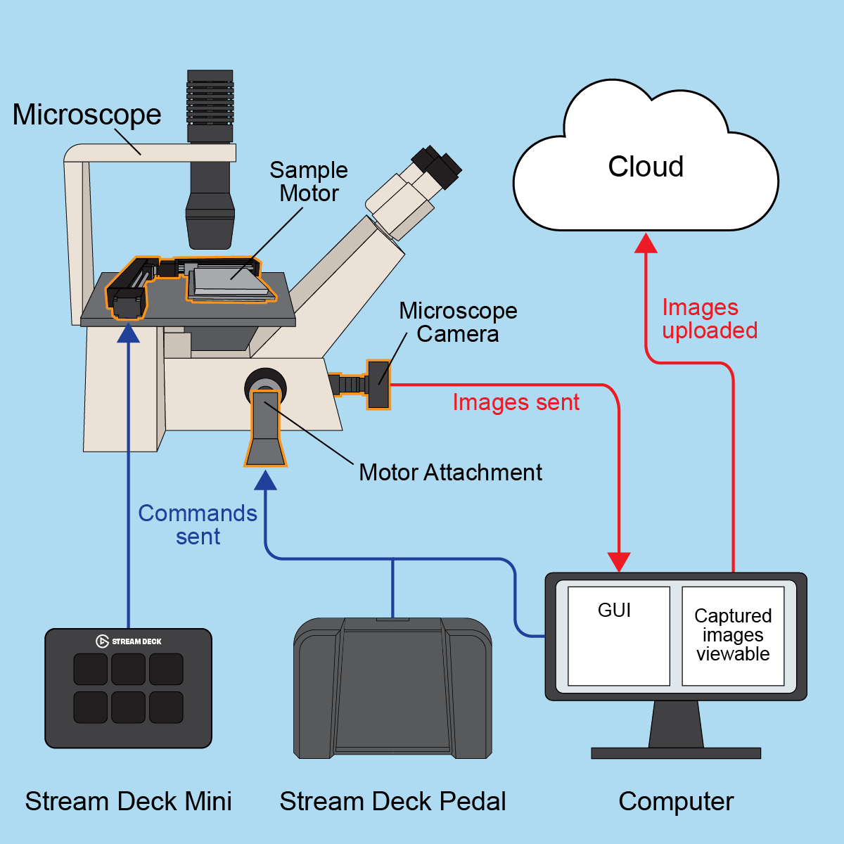
Final Graphical Abstract
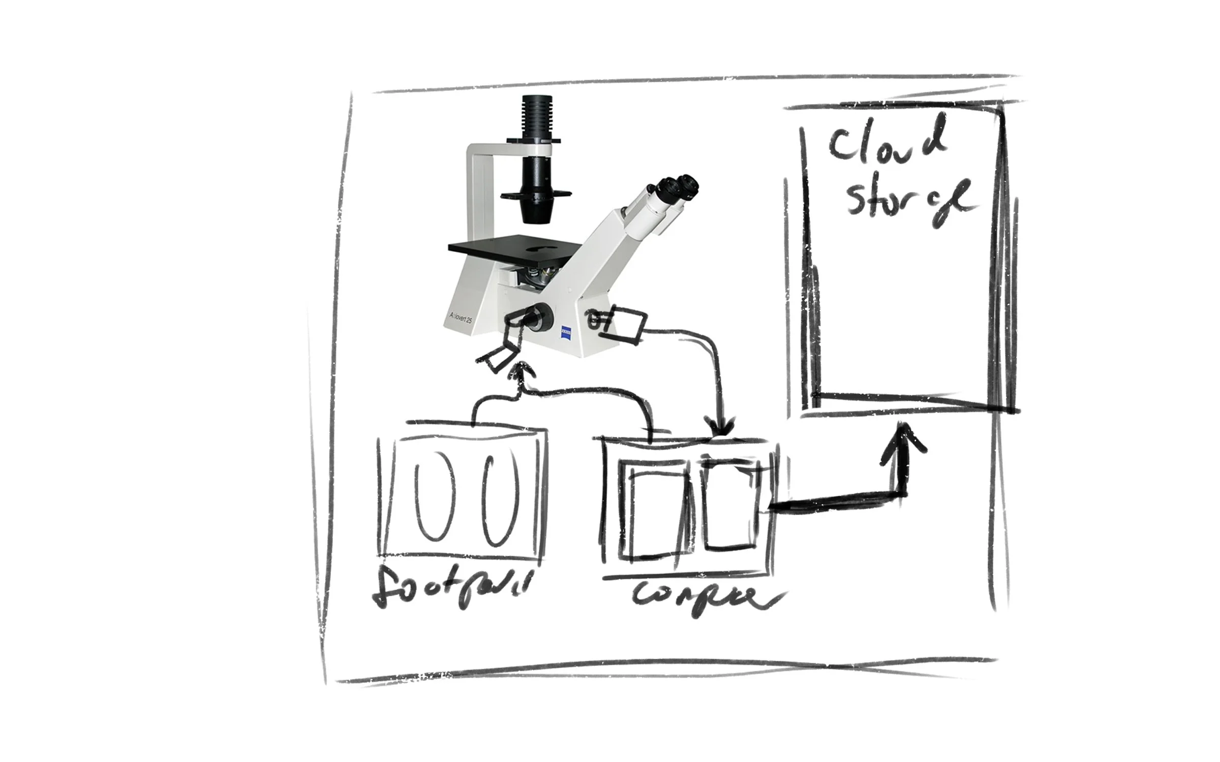
Initial sketch from first meeting with client
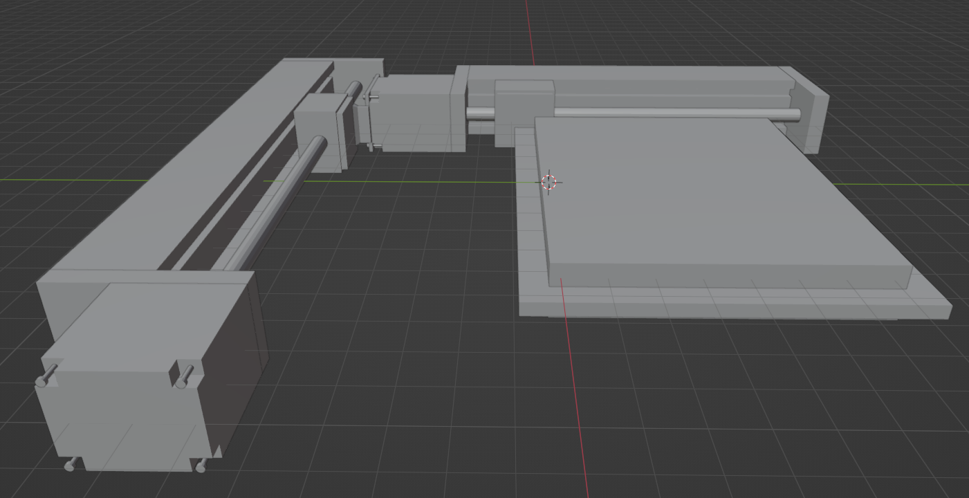
Recreated the hardware in Blender for accuracy and perspective

One of the first designs with color.
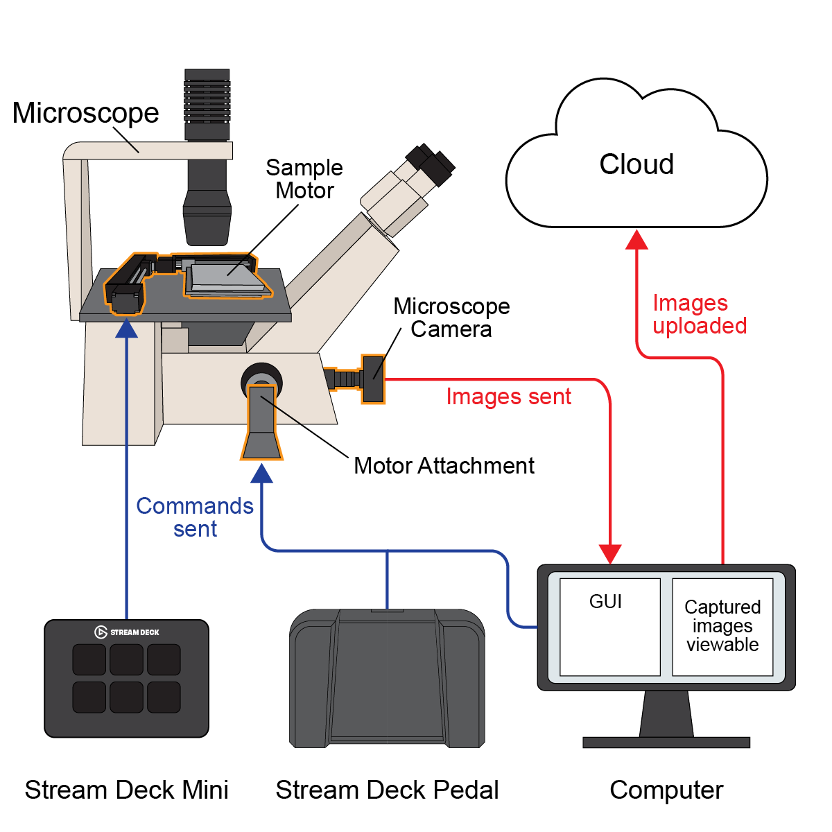
Graphics go through multiple iterations until we land on something that the team is happy with.